
Description:
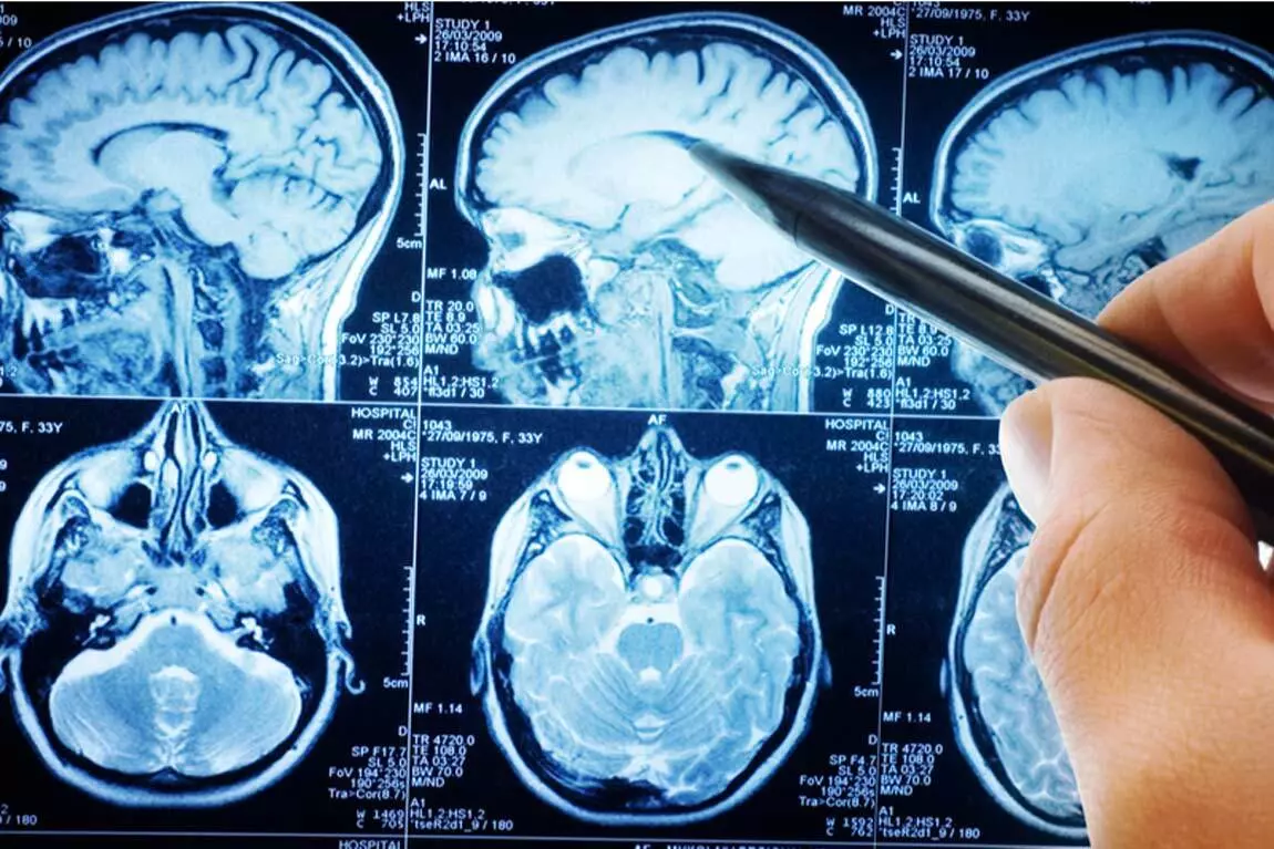
Neurosurgery is a branch of surgery that deals with the diagnosis, treatment, and management of disorders affecting the central nervous system (brain and spinal cord), peripheral nervous system, and supporting structures including the skull, spine, and cerebral blood vessels. Craniotomy for brain tumor resection is one of the most complex and delicate surgical procedures, in which a portion of the skull is temporarily removed to allow the surgeon to access the brain tissue and precisely remove the tumorous lesion. This procedure is indicated for primary brain tumors (such as meningioma, glioma, astrocytoma), metastatic brain tumors (spread from other parts of the body), and certain benign lesions that cause compressive symptoms or seizures. Patients typically present with intractable headaches, seizures (epilepsy), focal neurological deficits (such as limb weakness, speech or visual disturbances), personality or cognitive changes, nausea and vomiting, or in acute cases, decreased level of consciousness.
Treatment Options:
The main treatment modalities for brain tumors include:
Conservative / Observational Management – close monitoring with serial imaging for small, asymptomatic, or slow‑growing benign tumors
Medical Treatment – corticosteroids (for edema reduction), antiepileptic drugs (for seizure control), and chemotherapy or targeted therapy (for certain tumor types)
Radiotherapy – external beam radiation, stereotactic radiosurgery (Gamma Knife, CyberKnife) for inoperable or residual tumors
Craniotomy with Tumor Resection – surgical opening of the skull to remove the tumor (total or subtotal resection)
Biopsy – stereotactic or open biopsy for histological diagnosis without tumor removal
Endoscopic / Minimally Invasive Surgery – keyhole approaches or neuroendoscopy for selected deep‑seated or intraventricular tumors
Awake Craniotomy – surgery performed with the patient awake during part of the procedure to monitor and preserve critical brain functions (speech, motor, sensory areas)
Combined Modality Therapy – surgery followed by radiotherapy and/or chemotherapy as part of a multidisciplinary approach
Procedures:
Craniotomy for brain tumor resection is performed under general anesthesia and typically takes 120 to 300 minutes (2 to 5 hours), depending on the size, location, and complexity of the tumor, as well as the need for intraoperative monitoring. The procedure begins with the patient positioned on the operating table with the head fixed in a three‑pin head holder (Mayfield clamp) to ensure absolute stability. The surgical site is shaved and prepared. A curved or U‑shaped incision is made in the scalp, and the skin and muscle layers are reflected to expose the underlying skull. One or more burr holes are drilled, and a bone flap is created using a high‑speed craniotome. The bone flap is carefully removed and set aside for later reattachment. The dura mater (thick membrane covering the brain) is opened, and the tumor is identified using neuronavigation, intraoperative ultrasound, or MRI guidance. Using microsurgical techniques and an operating microscope, the tumor is dissected and resected with meticulous care to preserve surrounding healthy brain tissue and critical neural structures. Intraoperative neurophysiological monitoring (such as motor evoked potentials, somatosensory evoked potentials, and electrocorticography) may be used to map and protect functional areas of the brain. In cases of eloquent cortex involvement, awake craniotomy with intraoperative mapping is performed. After complete or subtotal tumor removal, meticulous hemostasis is achieved. The dura is closed in a watertight fashion, the bone flap is reattached using mini‑plates and screws, and the scalp is closed in layers. A drain is sometimes placed to prevent fluid accumulation. The patient is transferred to the intensive care unit (ICU) or neurosurgical ward for close postoperative monitoring.
This package includes:
- Preoperative consultation with neurosurgeon, neurologist, anesthesiologist, and intensivist
- Complete laboratory workup (CBC, coagulation profile, liver/kidney function, electrolytes, blood glucose, and tumor markers if indicated)
- Advanced neuroimaging – MRI brain with contrast, MR spectroscopy, functional MRI (fMRI), and diffusion tensor imaging (DTI) for eloquent area mapping (if indicated)
- CT angiography or digital subtraction angiography (DSA) – for assessment of vascular anatomy (if indicated)
- Electroencephalogram (EEG) – if seizures are present
- Preoperative neurological assessment and functional status evaluation
- Stereotactic neuronavigation system (for intraoperative tumor localization and guidance)
- Craniotomy with microsurgical tumor resection under general anesthesia (with or without awake technique)
- Intraoperative neurophysiological monitoring (motor evoked potentials, somatosensory evoked potentials, electrocorticography) – if indicated
- Operating microscope and advanced microsurgical instruments
- One to three days ICU admission for close monitoring (depending on case complexity)
- Two to seven days hospital admission (total stay, depending on recovery)
- Postoperative pain management, anti‑edema therapy (corticosteroids), and antiepileptic prophylaxis
- Wound care and drain management
- Postoperative neurological assessment and monitoring
- Discharge medications (antiepileptics, corticosteroids, analgesics) and comprehensive follow‑up plan
- Histopathology report with tumor typing, grading, and molecular markers (results within 7‑14 days)
- One follow‑up appointment at 2‑4 weeks post‑discharge with MRI for outcome assessment
- Preoperative medical clearance beyond standard evaluation (e.g., cardiology consult, stress testing, echocardiography) – if required, costs are separate
- Extended ICU stay beyond three days due to complications (e.g., cerebral edema, hemorrhage, infection, seizures, hydrocephalus)
- Extended hospital stay beyond seven days due to complications
- Radiotherapy (external beam radiation or stereotactic radiosurgery) – if required postoperatively, costs are separate
- Chemotherapy or targeted therapy – if required postoperatively, costs are separate
- Second‑look surgery or reoperation for recurrence or complications
- Consultation with other specialists (e.g., oncologist, radiation oncologist, psychiatrist) if additional treatment is required
- Rehabilitation services (physical therapy, occupational therapy, speech therapy) beyond initial inpatient consultation
- Prosthetic materials (such as custom cranioplasty implants for bone reconstruction in cases of bone involvement)
- Blood transfusion (if required – costs are separate)
- Home nursing care or home rehabilitation services after discharge
Instructions (Preparation):
Do not eat or drink anything (except essential medications with a small sip of water) for at least 6‑8 hours prior to surgery (general anesthesia protocol).
Bring all your previous medical records, imaging reports (MRI, CT scans), histopathology results (if biopsy was previously performed), and prior treatment history.
Inform your neurosurgeon and anesthesiologist about any history of bleeding disorders, allergies, current medications (especially anticoagulants, antiplatelets, antiepileptics, or corticosteroids), and any underlying medical conditions (diabetes, hypertension, cardiac, pulmonary, or renal disease).
If you are on anticoagulant or antiplatelet therapy (e.g., aspirin, warfarin, clopidogrel), discuss with your surgeon when to stop these medications (typically 5‑7 days prior to surgery).
If you have a history of seizures, ensure your antiepileptic medication is taken as scheduled.
If you are on steroids (for brain edema), continue as directed by your surgeon.
If you are a smoker, stop smoking at least 2‑4 weeks before surgery to reduce anesthetic risks and improve wound healing.
Arrange for a family member or caregiver to assist you during the first few weeks after discharge.
Terms and Conditions:
All neurosurgical procedures are performed using the most advanced surgical techniques, including stereotactic neuronavigation, intraoperative MRI/CT (if available), operating microscopes, and intraoperative neurophysiological monitoring to ensure maximum safety, precision, and preservation of neurological function. The surgical team consists of highly experienced neurosurgeons, neuroanesthesiologists, and specialized intensive care staff. Strict aseptic techniques and infection control protocols are followed to minimize complications. The procedure is tailored to each patient based on tumor type, location, size, and functional risk.
Patient Eligibility:
All procedures are conducted with the most advanced equipment and techniques.
Appropriate candidates for craniotomy with tumor resection include:
Patients with confirmed brain tumor (primary or metastatic) on imaging and/or biopsy
Patients with progressive neurological deficits, intractable seizures, or symptoms of raised intracranial pressure
Patients with tumor accessibility for safe surgical resection (based on anatomical location and functional mapping)
Patients with good performance status (able to tolerate surgery and anesthesia)
Patients with tumors that are radio‑resistant or when histopathological diagnosis is essential
Contraindications (High‑Risk Patients):
Patients with the following conditions are considered high‑risk and require thorough preoperative multidisciplinary assessment:
Extremely poor general condition (severely impaired performance status) with inability to tolerate general anesthesia
Uncontrolled intracranial pressure with impending herniation (requires urgent intervention)
Extensive deep‑seated or infiltrative tumors involving bilateral hemispheres, brainstem, or both thalami with no safe surgical corridor
Severe coagulopathy or bleeding diathesis not correctable with medical therapy
Active systemic infection or sepsis
Uncontrolled medical conditions (uncontrolled diabetes, severe cardiac, pulmonary, or renal failure)
Age over 80 years with multiple comorbidities – requires careful risk‑benefit assessment
Multiple extensive intracranial metastases without surgical benefit in terms of functional outcome or survival
Contact:
Jam Hospital is available 24/7 for consultations, admissions, and emergency support. Our dedicated neurosurgical team is ready to answer your questions and guide you through the entire treatment journey.
Goals:
To provide the most effective, safe, and individualized surgical treatment to achieve maximal safe tumor resection, preserve neurological function, relieve symptoms, improve quality of life, and ensure comprehensive multidisciplinary care (surgery, neuro‑oncology, radiotherapy, and rehabilitation) for optimal long‑term outcomes and patient satisfaction.